Have you considered the impact of the menopause on bone health? Jacky Forsyth explains how to improve and maintain bone health in the menopausal years.
This week marked World Menopause Day and, for this important event, we wanted to share some technical insight into the effects of the menopause on bone health.
In the UK, one in two women over the age of 50 suffers from fractures caused by osteoporosis (porous bones)1, which can have devastating effects on a woman’s quality of life. Not only that, but the risk of mortality as a result of an osteoporotic hip fracture is greater for women aged 50 and over than it is from all cancers combined2. However, one of the most neglected aspects of general fitness is that of bone health, since the adaptation of the bone to exercise is not as obvious as, for instance, muscle adaptation. The risk of osteoporosis increases when a woman enters menopause and, because women spend almost a third of their lifetime in a menopausal state3, it is important to understand the risk factors that can make postmenopausal women more likely to suffer from this debilitating disease, as well as the exercise and dietary recommendations for improving bone health.
The purpose of this article is, therefore, to review menopausal-related osteoporosis and give recommendations for improving and maintaining bone health at menopause and beyond.
Bone fracture osteoporosis is a progressive disease characterised by low bone mass and deterioration in the bone microarchitectural structure, leading to an increased susceptibility for fracture. The morbidity and mortality associated with osteoporotic fracture incurs substantial social and economic cost, making osteoporosis a major public health concern4.
Osteoporosis is often described as a ‘silent disease’ as it is not possible to know whether someone is suffering from osteoporosis until they have their first bone fracture. By this time, it is usually only preventative measures that are possible. Common sites of osteoporotic fracture include the femoral neck of the hip, the distal radius of the forearm, and the vertebrae of the spine. The bone fracture itself can be devastating, especially in an older population, since it means a loss of independence, if not for the long term, but for the short term. There is also the risk of death, with mortality being greatest in the first few months to a year following fracture of the hip or vertebrae5. As well as the debilitating effects and increased mortality risk of fracture, there is also a strong association between a fragility (osteoporotic) fracture in menopausal females and other disease states, especially cardiovascular disease6. Although the prognosis for osteoporosis can be good (with appropriate management), it is much better to try and avoid osteoporosis in the first place, rather than reacting to its consequences. To avoid osteoporosis, it is pertinent, especially for menopausal women, to consider the risk factors that increase susceptibility.
Risk groups
Non-modifiable risk factors for osteoporosis include genetics (having a family history of osteoporosis), age (being older), sex (being female), and race (there is a greater risk in Caucasian and Asian females)7. Modifiable risk factors for women include: a deficiency in calcium and vitamin D; having a body mass index of ≤19kg/m2; chronic smoking (>15 cigarettes per day); lack of physical activity; excessive alcohol consumption (more than the recommended 14 units of alcohol per week); gastro-intestinal disorders (due to a lack of absorption of proteins and key vitamins/minerals); glucocorticoid use; and anything that reduces oestrogen, including the menopause, amenorrhoea (absence of a normal menstrual cycle for three months or more) and early hysterectomy8. For the postmenopausal female, because of the already increased risk of osteoporosis, it is important to check bone health if these other risk factors are, or have been, present.
One of the most widely used methods of assessing bone health is through DXA (dual-energy X-ray absorptiometry) scanning. The scan measures bone mineral density (BMD) and uses T-scores to measure how much a person’s BMD deviates from that of a sex-matched, young, healthy adult.
In postmenopausal females, osteoporosis is defined as a BMD T-score that is -2.5 or lower, and osteopaenia (low bone health) as a BMD T-score that is between -1.0 and -2.48. A DXA scan is offered through the NHS for menopausal women if they suffer from a fracture after a minor, low-trauma fall or injury, and for those who have one or more of the risk factors described previously9.
The prevalence of osteoporosis and associated fracture increases substantially when women enter the menopause10. The reason for this increased risk is due to the associated decline in the hormone oestrogen. Specifically, oestrogen plays a role in bone remodelling (the normal process of bone turnover that occurs throughout life), and the process involves the family of tumour necrosis factor (TNF) cells: RANKL (the name is derived from Receptor Activator of Necrosis factor-Kappa B Ligand), RANK, and osteoprotegerin (OPG)11. When RANKL binds with RANK, osteoclasts (the bone-absorbing cells) are activated, leading to increased osteoclastic activity; in other words, bone is broken down. Osteoprotegerin works as a decoy receptor, which means that it prevents the binding of RANKL to RANK. When oestrogen levels are optimal, RANKL expression by osteoblasts is inhibited and OPG blocks the binding of RANKL to RANK; hence, osteoclastic activity is reduced – less bone is broken down. When oestrogen levels are low, however, there is both an increased expression of RANKL and a decrease in OPG to block the RANKL and RANK binding12. As a result, osteoclastic activity outstrips the pace of osteoblast activity (bone building), resulting in net bone loss13. In addition, increases in the follicle-stimulating hormone (FSH), which occurs during the peri-menopausal period, increases RANKL expression14 and hence contributes to bone loss.
Exercise as medicine
It has long been known that exercise improves bone health. In the 1960s’ space age, it was found that astronauts were returning to Earth having weakened bone structures. The mechanism by which bone adapts to exercise is described by the mechanostat theory15, which essentially postulates that bone turnover (growth and loss) is stimulated by local mechanical loading (i.e., exercise) through a series of cell signalling processes. The mechanostat theory explains why athletes who take part in non-weight-bearing sports, such as swimming and cycling, have a much lower BMD than those who take part in weight-bearing sports16, and why tennis players have greater BMD in their playing arm than their non-playing arm17. Bone response to exercise is, therefore, site and load specific.
When oestrogen levels are suboptimal, the type of exercise that is required needs to be as targeted as possible. For instance, among menopausal women, some exercise interventions for improving bone health have not always been effective18 since the exercise has not been of the right loading, ‘oddness’ or impact18. For the lower body, jumping has been shown to be particularly useful, especially for improving hip BMD19. In contrast, walking and jogging bring about only modest improvements in bone health20, possibly because the bone adapts quickly to the repetitious nature of this low load. For the postmenopausal female, therefore, small bouts of jumping (i.e., 10 jumps daily, with 10-second rest intervals) should be recommended to improve lower-body BMD21. The height of the jump should be determined by the capability of the individual, but jumping should be carried out bare-footed on a hard surface.
Most research interventions have focused on bone-targeted exercise for the lower body. Since bone health is site specific, and because a distal forearm fracture is common, it is important to also target the upper body. Based on a systematic review of 28 studies (unpublished data from the author), it seems that high force, resistance training of the upper body is effective for improving upper-body BMD, especially for postmenopausal women. These resistance training interventions consisted of between two and three sets of eight to 12 repetitions at around 80% 1RM, with sessions completed twice a week – so probably much higher than might typically be prescribed for an older population. As well as resistance training, there were two research studies that looked at impacting the bones of the hand and wrist directly, through having participants drop onto a wall with an outstretched arm22. The total number of these ‘wall drops’ was progressed to 40 per session, with two to four sessions completed each week. For the upper body, therefore, postmenopausal females need high bone-loading exercise for there to be an effect on BMD, although more research is needed to determine the precise mode, frequency and type of exercise that is required. In summary, for postmenopausal women, bone-targeted, multi-component exercise, such as muscle-strengthening exercise, and exercise that is dynamic, of high impact, discrete (with rest bouts) and unusual, with all areas of the body targeted, should be recommended.
Foods to help

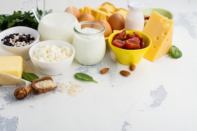
Ensuring optimal nutrition is an important strategy for reducing the risk of osteoporosis1. The foods most associated with an improvement in bone health include products rich in calcium, protein and vitamin D, as well as potassium, phosphorus, magnesium and zinc23. Good sources of calcium are found in things such as dairy products, green leafy vegetables and dried fruit. Supplementation with calcium should be avoided, due to the association that has been found with heart disease 24. As well as ensuring good quantities of sunlight exposure25, good food sources of vitamin D include oily fish, mushrooms, and some fortified dairy products. The recommended dietary intake for women over the age of 50 is ≥1000mg/day for calcium, 800 IU for vitamin D, and 1g/kg bodyweight for protein26. Improved balance in micronutrients can be achieved through consumption of pulses, vegetables, whole cereals and nuts. One lesser-known food product that has been found to improve bone health in postmenopausal women is dried plums, also known as prunes27. Prunes, being rich in polyphenolic compounds, inhibit RANKL expression by osteoclasts, which decrease bone resorption (breakdown)28. Prunes also contain boron, which stabilises and extends the half-life of vitamin D, improves oestrogen availability, and reduces calcium loss29. The vitamin K in the prunes also influences bone health by helping to improve calcium balance27. For peri-menopausal and menopausal women, supplementing the diet with six to eight prunes per day while undertaking dynamic, high-impact exercise would seem to be an effective lifestyle strategy for increasing or maintaining bone health.
Summary
Because the number of women over 50 years of age in the UK is projected to increase30, most women will be spending a large proportion of their lifetime beyond the menopausal transition. The symptoms that are typically associated with the menopause include hot flushes, sleep disturbance, headaches, vaginal dryness, reduced libido, anxiety and depression31, but one of the more debilitating and life-threatening outcomes of menopause is that of osteoporosis. As highlighted in this article, strategies to improve and maintain bone health for postmenopausal women include exercise that is sufficiently intense (but of a very short duration with rests inserted) to evoke bone growth, at the same time as eating a diet rich in essential nutrients, gained from products such as dairy, green leafy vegetables, oily fish and prunes.
About the author
Dr Jacky Forsyth is an associate professor in exercise physiology at Staffordshire University. Her research is in the area of women’s exercise and health. Jacky is vice chair of the Women in Sport and Exercise Academic Network (WiSEAN), the purpose of which is to grow, strengthen and promote research on women in sport and exercise.
@JackyForsyth
@WISE_AN
References
- Weaver, C.M., Gordon, C.M., Janz, K.F., Kalkwarf, H.J., Lappe, J.M., Lewis, R., Zemel, B.S. (2016) The National Osteoporosis Foundation’s position statement on peak bone mass.
- Cummings, S.R., Black, D.M. and Rubin, S.M. (1989) Lifetime risks of hip, Colles’, or vertebral fracture and coronary heart disease among white postmenopausal women, Archives of Internal Medicine, 149(11), 2445-2448.
- Ji, M.-X. and Yu, Q. (2015) Primary osteoporosis in postmenopausal women, Chronic Diseases and Translational Medicine, 1(1), 9-13, http://doi.org/10.1016/j.cdtm.2015.02.006
- Vasikaran, S., Cooper, C., Eastell, R., Griesmacher, A., Morris, H.A., Trenti, T. and Kanis, J.A. (2011) International Osteoporosis Foundation and International Federation of Clinical Chemistry and Laboratory Medicine Position on bone marker standards in osteoporosis, Clinical Chemistry and Laboratory Medicine, 49(8), https://doi.org/10.1515/CCLM.2011.602
- Leboime, A., Confavreux, C.B., Mehsen, N., Paccou, J., David, C. and Roux, C. (2010) Osteoporosis and mortality, Joint Bone Spine, 77, S107–S112, http://doi.org/10.1016/S1297-319X(10)70004-X
- Lello, S., Capozzi, A. and Scambia, G. (2017) Osteoporosis and cardiovascular disease, Maturitas, 100, 103, http://doi.org/10.1016/j.maturitas.2017.03.039
- Forsyth, J.J. and Davey, R.C. (2008) Bone health, Exercise Physiology in Special Populations, http://doi.org/10.1016/B978-0-443-10343-8.00007-X
- Kanis, J.A. and Kanis, J.A. (1994) Assessment of fracture risk and its application to screening for postmenopausal osteoporosis: Synopsis of a WHO report, Osteoporosis International, 4(6), 368-381, http://doi.org/10.1007/BF01622200
- NHS Choices (2016).
- Boschitsch, E.P., Durchschlag, E. and Dimai, H.P. (2017) Age-related prevalence of osteoporosis and fragility fractures: real-world data from an Austrian Menopause and Osteoporosis Clinic, Climacteric, 20(2), 157-163, https://doi.org/10.1080/13697137.2017.1282452
- Scheurer, H. (2013) Osteoblasts: Morphology, functions and clinical implications: Morphology, functions and clinical implications,Hauppauge: Nova.
- Schett, G. (2011) Effects of inflammatory and anti-inflammatory cytokines on the bone, European Journal of Clinical Investigation, 41(12), 1361-1366, http://doi.org/10.1111/j.1365-2362.2011.02545.x
- Marques, E.A., Mota, J., Viana, J.L., Tuna, D., Figueiredo, P., Guimarães, J.T. and Carvalho, J. (2013) Response of bone mineral density, inflammatory cytokines, and biochemical bone markers to a 32-week combined loading exercise programme in older men and women, Archives of Gerontology and Geriatrics, 57(2), 226-233, http://doi.org/10.1016/j.archger.2013.03.014
- Colaianni, G., Cuscito, C. and Colucci, S. (2013) Review article FSH and TSH in the regulation of bone mass: The pituitary/immune/bone axis, Clinical and Developmental Immunology, http://doi.org/0.1155/2013/382698
- Frost, H.M. (1987) Bone mass and the mechanostat: A proposal, The Anatomical Record, 219(1), 1-9, http://doi.org/10.1002/ar.1092190104
- Hind, K., Gannon, L., Whatley, E., Cooke, C. and Truscott, J. (2012) Bone cross-sectional geometry in male runners, gymnasts, swimmers and non-athletic controls: a hip-structural analysis study, European Journal of Applied Physiology, 112(2), 535-541, http://doi.org/10.1007/s00421-011-2008-y
- Ducher, G., Tournaire, N., Meddahi-Pellé, A., Benhamou, C.L. and Courteix, D. (2006) Short-term and long-term site-specific effects of tennis playing on trabecular and cortical bone at the distal radius, Journal of Bone and Mineral Metabolism, 24(6), 484-490, http://doi.org/10.1007/s00774-006-0710-3
- Kelley, G.A. and Kelley, K.S. (2006) Exercise and bone mineral density at the femoral neck in postmenopausal women: A meta-analysis of controlled clinical trials with individual patient data, American Journal of Obstetrics and Gynecology, 194(3), 760-767, https://doi.org/10.1016/j.ajog.2005.09.006
- Babatunde, O. and Forsyth, J. (2013) Effects of lifestyle exercise on premenopausal bone health: a randomised controlled trial, Journal of Bone and Mineral Metabolism, 32(5), 563-572, http://doi.org/10.1007/s00774-013-0527-9
- Martyn-St James, M. and Carroll, S. (2008) Meta-analysis of walking for preservation of bone mineral density in postmenopausal women, Bone, 43(3), 521-531, http://doi.org/10.1016/j.bone.2008.05.012
- Babatunde, O., Forsyth, J.J. and Gidlow, C.J. (2012) A meta-analysis of brief high-impact exercises for enhancing bone health in premenopausal women, Osteoporosis International, 23(1), 109-119, http://doi.org/10.1007/s00198-011-1801-0
- Greenway, K.G., Walkley, J.W. and Rich, P.A. (2015) Impact exercise and bone density in premenopausal women with below average bone density for age, European Journal of Applied Physiology, 115(11), 2457-2469, http://doi.org/10.1007/s00421-015-3225-6
- Rizzoli, R., Bischoff-Ferrari, H., Dawson-Hughes, B. and Weaver, C. (2014) Nutrition and Bone Health in Women after the Menopause, Women’s Health, 10(6), 599-608, http://doi.org/10.2217/WHE.14.40
- Reid, I.R. (2014) Should We Prescribe Calcium Supplements For Osteoporosis Prevention? Journal of Bone Metabolism, 21(1), 21, http://doi.org/10.11005/jbm.2014.21.1.21
- Melin, A., Wilske, J., Ringertz, H. and Sääf, M. (2001) Seasonal variations in serum levels of 25-hydroxyvitamin D and parathyroid hormone but no detectable change in femoral neck bone density in an older population with regular outdoor exposure, Journal of the American Geriatrics Society, 49(9), 1190-1196.
- Kanis, J.A., McCloskey, E.V., Johansson, H., Cooper, C., Rizzoli, R. and Reginster, J.-Y. (2013) European guidance for the diagnosis and management of osteoporosis in postmenopausal women, Osteoporosis International, 24(1), 23-57, http://doi.org/10.1007/s00198-012-2074-y
- Wallace, T.C. (2017) Dried plums, prunes and bone health: A comprehensive review, Nutrients, 9(4), http://doi.org/10.3390/nu9040401
- Hooshmand, S., Brisco, J.R.Y. and Arjmandi, B.H. (2014) The effect of dried plum on serum levels of receptor activator of NF-κB ligand, osteoprotegerin and sclerostin in osteopenic postmenopausal women: a randomised controlled trial, British Journal of Nutrition, 112(1), 55-60, http://doi.org/10.1017/S0007114514000671
- Pizzorno, L. (2015) Nothing boring about boron, Integrative Medicine (Encinitas, Calif.), 14(4), 35-48, retrieved from http://www.ncbi.nlm.nih.gov/pubmed/26770156
- Rutherford, T. (2012) Population ageing: statistic, retrieved from http://researchbriefings.files.parliament.uk/documents/SN03228/SN03228.pdf
- Bauld, R. and Brown, R.F. (2009) Stress, psychological distress, psychosocial factors, menopause symptoms and physical health in women, Maturitas, 62(2), 160-165, https://doi.org/10.1016/j.maturitas.2008.12.004


